Ultrasound Imaging
Cardiovascular Ultrasound
Cardiovascular ultrasound, also known as an echocardiogram or heart ultrasound, is a non-invasive imaging technique that uses sound waves to create images of the heart’s structure and function, aiding in the diagnosis and management of various heart conditions.
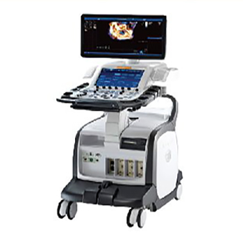
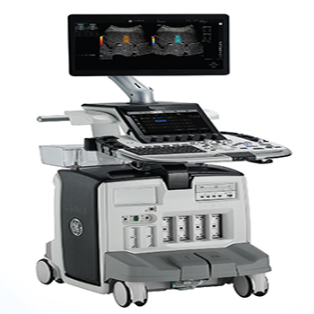
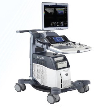
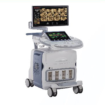
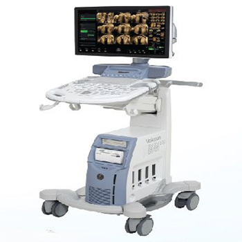
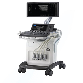
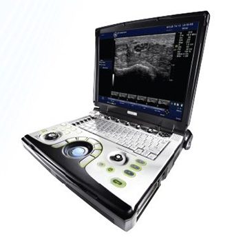
Vivid E95 (Ultra Edition)-Premium 4D Cardiac Ultrasound System
- High resolution 24′ wide LED monitor
- AFI ILV, RV, LA)
- 40 MVQ, AVQ, LVQ, RVQ, TVQ
- Auto measurement 2D
- Auto Spectrum Recognition
- Auto VQ, Flexi Light
- HD Colour, 40 Marker
- Anatomical M-mode both on line & offline
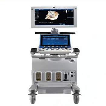
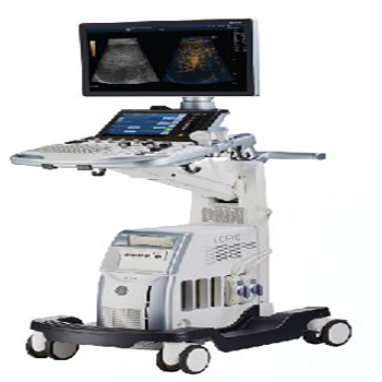
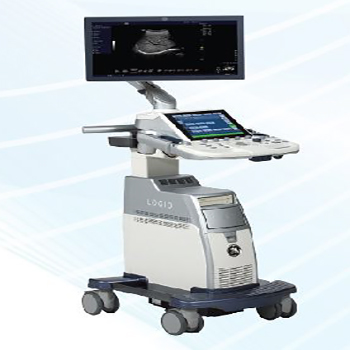
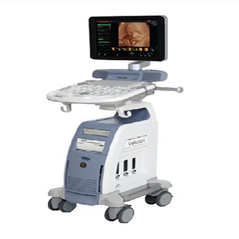
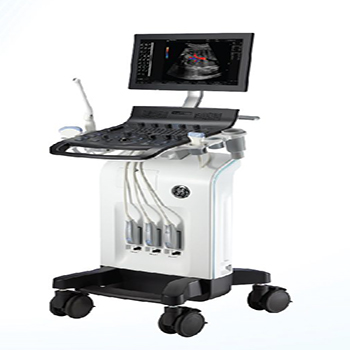
Vivid S70 (Ultra Edition)-Robust System With 4D TEE capability
- 21.5" wide screen HD display
- Ergonomic FlexFit design
- Auto measurement 2D
- AFI, Stress echo, TVI, TOI, TT. TSI
- Auto Spectrum Recognition
- 12" ultra-high-resolution wide screen format
- 40 TEE Capabilities Smart standby
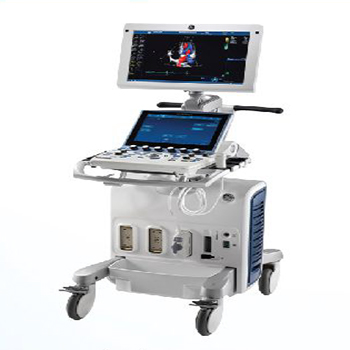
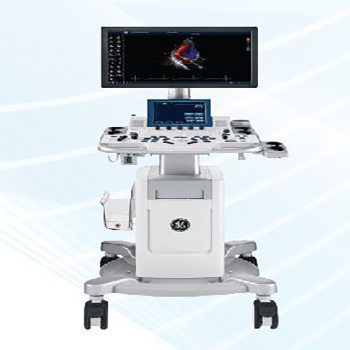
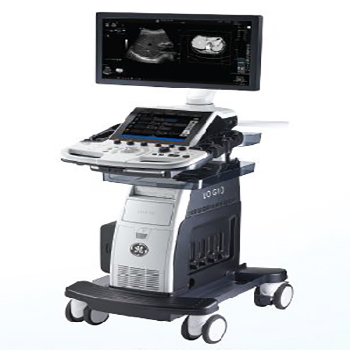
Vivid S60 (Ultra Edition)-Robust 2D System Takes Cardiovascular Ultrasound To New Heights
- 21.5" wide screen HD display
- 12" multi-touch LCD screen with tablet like performance
- Al Auto measurement 20
- Stress echo, TVI, TOI, Tl TSI
- Flex Fit - Adjust control panel and monitor
- Smart standby without AC power
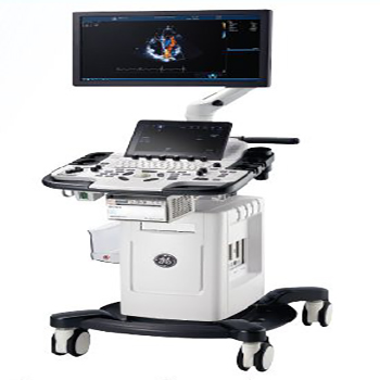
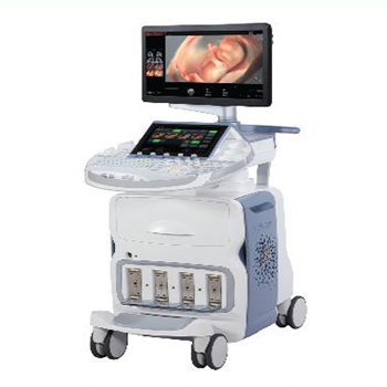
Vivid T9 (Ultra Edition)-Pat1ent Care.Elevated .
- 21.5" wide screen HD display
- Ergonomic FlexFit design
- Intuitive control layout includes the 10.Uil multi-touch screen
- Cardiac auto doppler- artificial intelligence-based tool
- AutoEF 2.0 - second-generation tool for assessing and quantifying ventricular wall motion
- Stress Echo
- AFI, Tl TVI, TOI. TSI, Q-Analysis
- Auto measure 20
- Articulating arm
Vivid T8 (Ultra Edition)-PoweredByAI.ElevatedByVou
- 21.5" wide screen HD display
- Intuitive control layout includes the 10.Uil multi-touch screen
- LVO Contrast, Tissue Tracking, Adv. Qscan Imaging, Q-Analysis
- Scan Coach; provides reminders/refresh information
- Smart Standby
- Tissue Velocity Imaging - captures dynamic information from moving heart tissue
- Blood Flow Imaging
- Stress Echo
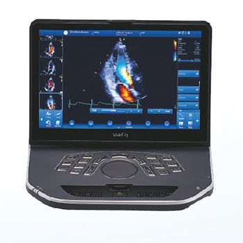
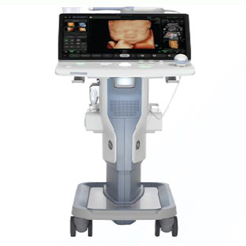
Vivid iq (Ultra Edition)-ThePawerToTakeVouPlaces.
- Light weight 5.2kg
- Al Auto measurement 2D
- AFI ILV, RV, LA}
- Stress Echo
- Auto EF, TT. TVI, TOI, TSI. Q-Analysis
- Auto Spectrum Recognition
- Closed surface design - allows cleaning with disinfectant solutions
General Imaging Ultrasound
General imaging ultrasound uses high-frequency sound waves to create real-time images of internal organs and tissues, aiding in the diagnosis and evaluation of various medical conditions without the use of ionizing radiation.
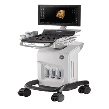
Logiq E10S-Empowering you to Make a Difference
- 22″ OLEO screen
- 12″ multi-touch screen
- UGAP, Micro-Vascular Imaging
- Auto Doppler Assist
- Start Assistant enables you to experience end-to-end workflow
- OB/Breost/Tyroid Tools for labelling, measuring and describing lesions, nodules, lymph nodes and parathyroid
- Acquire images in your preferred order or easily reorder images within the exam
- B-Steer + to enable enhanced visualization of the needles structure
- Multi-dimensional 2D sharewove Elostogrophy
- “Any-Plane” function for 3D and 4D data
Log iq S8-simply amazing
- 23″ LED screen
- 10.1″ multi touch screen
- S-Agile Architecture; dynamic image acquisition for all body types
- E-Series Transducers for improved sensitivity, penetration and image uniformity
- High-Definition Speckle Reduction Imaging (SRI-HD)
- CrossX Beam; combines multiple images into one clear appearance
- Coded Harmonic Imaging for enhanced near field resolution and far field penetration
- LOGIQ View facilitates excellent visualization and more clinical information.
- B-Flow Imaging enables direct visualization of blood flow, without the issues of doppler
Logiq S7-Simply Amazing
- 23″ LED screen
- 10.1″ multi touch screen
- S-Agile Architecture; dynamic image acquisition for all body types
- E-Series Transducers for improved sensitivity, penetration and image uniformity
- High-Definition Speckle Reduction Imaging (SRI-HD)
- CrossX Beam; combines multiple images into one clear appearance
- Coded Harmonic Imaging for enhanced near field resolution and far field penetration
- LOGIQ View facilitates excellent visualization and more clinical information.
- B-Flow Imaging enables direct visualization of blood flow, without the issues of doppler
Logiq P7-Makes It Easy For you
- Large 21.5″ monitor and accessible 10.4″ touchscreen
- Touch Control; easily adjust imaging parameters on touch panel
- Photo Assistant App; combine onotamicol photos and images in same report
- 2D Shear Wove Elostography – Quantitative estimate of tissue elasticity
- Remote Control App – Operate the system from on Android phone or tablet
- My Trainer software
- CRl,SRI, ATO, AO
Logiq P9-Make It Easy. Make It your own.
- Large 21.5″ monitor and accessible 10.4″ touchscreen
- Touch Control; easily adjust imaging parameters on touch panel
- Photo Assistant App; combine anatomical photos and images in same report
- HD Color – Sensitivity for visualizing small vessels and slow flow
- 2D Shear Wove Elostography – Quantitative estimate of tissue elasticity
- Remote Control App – Operate the system from on Android phone or tablet
- SonoDefense – Powerful data security features to help guard against costly breaches
- CRl,SRI, ATO, AO
Women’s Health Ultrasound
Women’s health ultrasound uses high-frequency sound waves to create images of internal organs and structures, particularly within the reproductive system, to diagnose and monitor various conditions, including pregnancy and gynecological issues.
Voluson E10-The Excellence You Demand The Standards You Set
- Radiantflow; delivers easy. fast visualization of even the tiniest of vessels
- SlowflowHD for blood perfusion visualizations
- HDlive™ technology suite helps easily obtain volume images with unprecedented depth and clarity
- HDRes elevates tissue differentiation. border definition and fine resolution eSTIC. e4D.
- eSnapshot to optimize Electronic 4D.
- SonoCNS helps properly align and display views and measurements of the fetal brain
- XDclear™ probe technology for achieving exceptional tissue and detail resolution
Voluson E8-Advanced Features. Simplified Work/low
- Radiantflow; delivers easy. fast visualization of even the tiniest of vessels
- SlowflowHD for blood perfusion visualizations
- Radiance System Architecture for simplified scanning and foster processing speeds
- Edison – Artificial Intelligence reduces keystrokes by 80% for foetal brain exams
- Proven interface coupled with automation and ergonomic design
- 4DView allows you to optimize. manipulate. and analyze volume ultrasound data offline
Voluson E6-Flexib1lity You Need Performance You Require
- SonoRenderlive facilitates 3D and 4D rendering
- XDclear™ probe technology for achieving exceptional tissue and detail resolution
- Uterine Trace for easy documentation of uterine shape
- HDlive™ technology suite helps easily obtain volume images with unprecedented depth and clarity
- SonoCNS helps properly align and display views and measurements of the foetal brain
Voluson S10 Expert-Your Expertise, Enhanced By Innovation
- Advanced VCI – Adjusts slice thickness to help enhance contrast resolution.
- Superb 2D and 3D/4D imaging. optimized for clarity and detail
- Vibrant visualization of anatomy ond function with advanced color Doppler
- HDlive™ technology suite; gets volume images with unprecedented depth and clarity
- Consistent image quality even in the most difficult to image patients
- SonoVCAD™heart for essential views of the foetal heart from a single STIC volume
- 3D Printing for rapid clinical prototyping. and parent bonding
- Advanced VCI -adjusts slice thickness to help enhance contrast resolution.
Voluson S8 Touch-Your Unique Demands Require Distinct Answers
- Excellent 2D and 3D/4D image quality
- HDlive™ technology for unprecedented depth. clarity and exceptional anatomical realism
- VCI with OmniView to easily view irregularly shaped structures
- Sano-automation technologies to decrease complexity and increase exam consistency
- Voluson xTouch; experience intuitive volume navigation on a 10.1″ touch panel
Voluson S8-Extraordmary V1s1on
- 23″ widescreen LED monitor
- Report preview – provides access to completed measurements and trending
- Adaptive control panel
- Battery Pack – provides up to 20 minutes of scan time
- Superb 2D and 3D image quality optimized for clarity and detail
- Color Doppler Quickly to quickly assess vascular anatomy and functions
- Achieve the penetration needed for all exam and body types
- HDlive™ technology for 3D and 4D imaging
- HDlive™ technology for exceptional anatomical realism
Voluson P8-Women's Health Imaging Simplified
- Excellent clarity and detail in 2D images
- Expand clinical confidence with 3D/4D technology
- Advanced Color Doppler for superb sensitivity into anatomy and function
- Consistent imaging even in difficult-to-image patients
- Sano-automation technologies
- SonoRenderlive facilitates 3D and 4D rendering
Voluson SWIFT+ -This Changes Everything
- 5″ High resolution full touch interface LED.
- Voluson core architecture. Light Weight and manoeuvrable.
- Sonolyst Improve quality and simplify fetal Anatomy Scans.
- Sano FHR for auto fetal heart rate measurement.
- Son biometry I HC. BPD. AC. FL. HL. CM Vp and cerebellum I Auto measurement.
- Uterine trace to obtain coronal plane of uterus.
- SonoCNS helps properly align and display views and measurements of the foetal brain.
- Battery Backup.
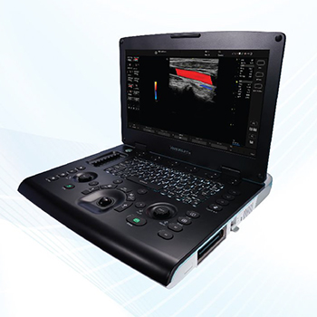
Primary Care Ultrasound
Primary care ultrasound, also known as point-of-care ultrasound (POCUS), is a non-invasive imaging technique used by primary care physicians to quickly assess and diagnose various conditions at the point of care, improving efficiency and patient satisfaction.
Versana Balance-care with canfidence
- S” LED Display
- CrossXBeam and SRI-HD to define structure borders clearly
- Height-adjustable console
- Broad range of probes
- Multiple ports to support a wide variety of exams
- Make automatic 2D measurements
- Scan Coach; 3D animation. illustrations and reference images to find the correct scan plane
- Voice comments; capture recorded voice comments overlaid on images Myocardial Doppler for imaging with colour overlay on the tissue image
Versana Premier-Powerful. Versatile. Productive.
- S” LED Display with 9.9″ touch screen
- Height adjustable console
- Gel wormer
- Wizz function for easy image quality optimization
- Needle Recognition clarifies the precise location of the needle point Tricefy Uplink; send images to the Tricefy cloud wirelessly for consultation TruScan to review and analyse images
- Myocardial Doppler for imaging with colour overlay on the tissue image Tomographic Ultrasound Imaging (TUI) to assess slices within a volume
Versana Active (Portable)-Advanced. Capable. Adaptable.
- Lightweight (5 KG with built in removable battery)
- New image processor with faster frame rates and updated algorithms Follow-Up Tool to compare the current and prior exams side-by-side Wizz function for easy image quality optimization
- Scan Assistant- lets you create standardized exam protocols
- Scan Coach – contextual reference tool for scan plane acquisition
Versa no Essentia I-Easy To use. Easy To Own.
- Automated measurement af IMT
- Wizz function far easy image quality optimization
- Small footprint and lightweight
- Simple and fast to use with automated calculations
- 3″ 16:9 LED Monitor
- 3 active probe connectors
- Color Doppler
- Scan Coach – real-time reference information ta help locate the correct scan plane
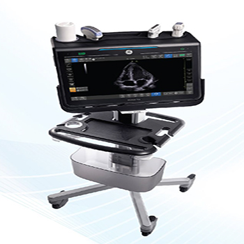
Point of Care Ultrasound
Point-of-care ultrasound (POCUS) is the use of ultrasound technology by clinicians at the patient’s bedside, rather than in a separate radiology department, to rapidly diagnose and guide treatment, augmenting physical examinations and expediting clinical decision-making.
Venue 40 (Portable)-surpnsmgly Easy To use
- PDI highlights vascular sensitivity
- PDI quantification far diagnosis and monitoring
- Choose from three probes – always connected – far quick access
- pre-configured application settings
- Intuitive touch interface
- Respond fast with quick boot-up
- Adjustable height and screen
Log iq e (Porta ble)-see Clearly. See Quickly. Guide Precisely.
- Optimized for head and neck. permitting right-out-of-the-box imaging
- Probe versatility – use a single probe for many applications
- Simple documentation; one click sends images to multiple storage locations
- Compact and battery-operated. easily move from patient to patient
- LOGIQ e pre-sets help simplify the selection of the right system settings
- for each application
- Precise visualization of law and slow blood flaw and accurate imaging of vascularity
Vscan Access (Handheld)-Assessing Risk. Expanding Reach.
- Intuitive touchscreen
- Gesture-based control
- Clinical protocols and automated controls
- Damage-resistant screen
- Drop-tested dust-proof exterior
- Battery-powered
- Lightweight for portability
- Scan Coach tool to help optimize scan plane and probe position
- Growth tracking software to display foetal development over time
- Bluetooth® for wireless data transfer and mHealth applications