Diagnostic Imaging
Magnetic Resonance Imaging (MRI)
Magnetic Resonance Imaging (MRI) is a medical imaging technique that uses a strong magnetic field and radio waves to create detailed images of the inside of the body, without using ionizing radiation.
SIGNA Pioneer 3.0 T-Env1s1on What You've Always Wished MR Could Do
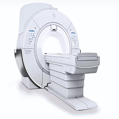
- Award winning AIR Coils Technology
- Free Breathing exams for Body MR with Auto Navigator
- SilentSuite -Silent MRI scan delivers unique patient comfort
- DISCO -Advanced MR technique to evaluate tumours activity and morphology
- 50% less power than conventional 3.0T wide bore MRI with UHE gradients sytem
- MAGiC: Multiple image contrasts in a single acquisition for compromised Patients Best in Class “GE” Magnet with superb homogeneity for high-quality off-centre MRI
- AIR Recon DL: deep-learning based reconstruction algorithm for exceptional Image
Signa Architect 3.0 T-Make The Unimaginable The Expected
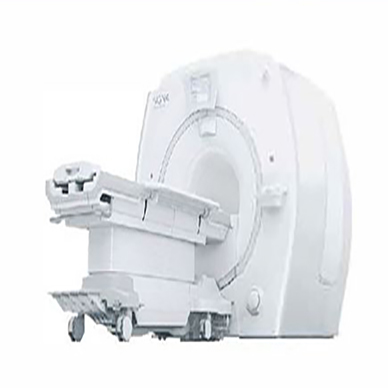
- Award winning AIR Coils Technology
- express table. with a memory foam surface
- Upto 50% faster acquisition time with Hyperworks
- Based on the SIGNA™Works platform to redefine productivity
- 48 Channel Head Coil and Coil Suite delivers phenomenal performance
- Total Digital Imaging ITDII feature brings startling advances in imaging
- Pre-loaded SIGNA™Works standard applications with SIGNA™ Architect DISCO -Advanced MR technique to evaluate tumours activity and morphology
SIGNA Explorer 1.5 T-Outstanding 1.5 T Performance
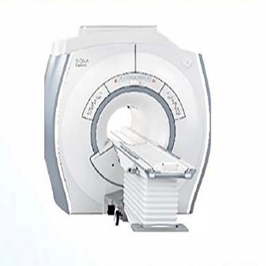
- Quality in the fastest scan times – The new standard for MR
- SilentSuite -Silent MRI scon delivers unique patient comfort
- 105 cm open flared Magnet and feet first MRI for patient comfort
- 34% less power than conventional 1.ST MRI with UHE gradient & RF system
- MAGiC: Multiple image contrasts in a single acquisition for compromised Patients
- Best in Class “GE” Magnet with superb homogeneity for high-quality off-centre MRI
- AIR Recon DL: deep-learning based reconstruction algorithm for exceptional Image
- Advanced acceleration techniques – Up to 50% foster acquisition time with Hyperworks
SIG NA Creator 1.5 T-Propel Your Imaging Abilities Into The Future
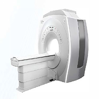
- Whole Body MRI with Scan Coverage 194 cm.
- Free Breathing exams for Body MR with Auto Navigator
- 105 cm open flared Magnet and feet first MRI for patient comfort
- Comes with GE post processing workstation-AW for advanced MRI
- Total Digital Imaging ITDI) feature brings startling advances in imaging
- 34% less power than conventional l.ST MRI with UHE gradient & RF system
- DISCO -Advanced MR technique to evaluate tumors activity and morphology.
- Best in Class “GE” Magnet with superb homogeneity for high-quality off-centre MRI
Signa Voyger 1.5 T-Skyrocket Yaur MR Performance
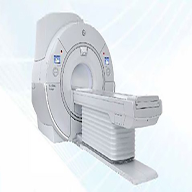
- Free Breathing exams for Body MR with Auto Navigator
- Quality in the fastest scan times – The new standard for MR
- SilentSuite -Silent MRI scan delivers unique patient comfort
- Best in Class Magnet homogeneity in a wide bore system MRI
- DISCO -Advanced MR technique to evaluate tumors activity and morphology
- AutoFlow suite of features makes workflow easier and more efficient than ever
- MAGiC: Multiple image contrasts in a single acquisition for compromised Patients.
- AIR Recon DL: deep-learning based reconstruction algorithm for exceptional Image
AIR Coil Technology-simple Better MR Coil
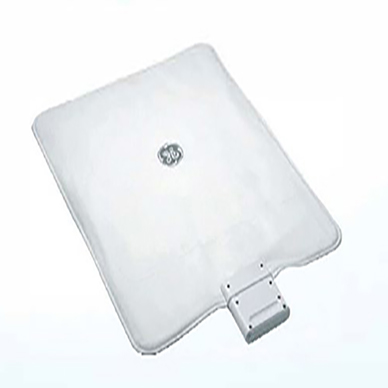
- 360 degrees of coverage
- Weighs upto 0.3 grams per cmZ
- Lightweight upto 60% lighter or more
- Automatically detect anatomy & prescribe slices with Al Rx™
- Industry leading versatality with simple more durable design
- Streamline & optimize scan setup with AIR Touch™
Computed Tomography (CT)
Computed Tomography (CT), also known as a CAT scan, is a medical imaging technique that uses X-rays to create detailed cross-sectional images of the body, allowing doctors to visualize internal organs, bones, and soft tissues.
Revolution ACTS ES-Your Aspirations. Realized
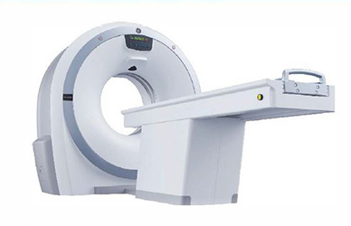
- Compact Geometry, compact design, less footprint and low power consumption
- Clarity panel detector, delivers 18 Ip/cm exceptional spatial resolution
- Sub-mm isotropic imaging for accurate detection
- All routine scans from head to toe
- Advanced clinical applications like peripheral angiography
Revolution ACTS EX-Your Aspirations. Realtzed
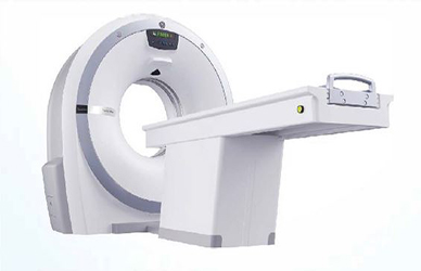
- Advanced imaging with 16 slices
- Clarity panel detector, delivers 18 Ip/cm exceptional spatial resolution
- Sub-millimetre scanning possible, all while maintaining a high dose efficiency
- Smart dose reduction technologies for patients – ASiR and ODM
- Quick and accurate scans with Sub sec scan, IQ enhancement with pitch booster
- Advanced applications including CT angiography, perfusion imaging, virtual endoscopy
Revolution ACTS Ex ert Edition-Elevate Your Radiology Practice

- Advanced imaging with 32 slices
- Clarity panel detector, delivers 18 Ip/cm exceptional spatial resolution
- ASiR™ provides breakthrough image quality with dose reduction upto 40%
- Virtual Endoscopy to visualize intraluminal structures
- Quick and accurate scans with Sub sec scan, IQ enhancement with pitch booster
- Neuro perfusion with advanced neuro package
- Advanced Clinical Applications and Technologies with AW workstation
Revolution ACT-One Act. Enhance Your Impact
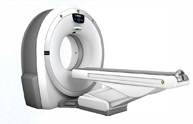
- Advanced imaging with 50 slices
- Clarity panel detector, delivers 18 Ip/cm exceptional spatial resolution
- High coverage of 35 mm/sec
- ASiR™ provides breakthrough image quality with dose reduction upto 40%
- Neuro perfusion, virtual endoscopy, angiographic studies
- Advanced Clinical Applications and Technologies with AW workstation
Revolution EVO-oefying Ttme
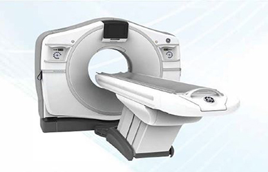
- 500 slice equivalent system with free breathe scan
- Clority panel detector with enhanced spatial resolution of 0.28 mm
- Upto 82% dose reduction using ASIR-V
- Unique Snapshot Freeze freezes coronary motion with a temporal resolution of 29 msec
- 5 beat cardiac acquisition for any heart rate
- Single acquisition SmartMAR for artifact-less imaging
- Elevate CT imaging with advanced applications in multimodality workstation
Revolution Maxima-overall.A Top Performer
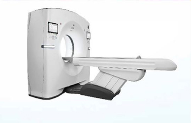
- Aunt Minee Europe Award winner for the best new r adiology device 2020
- Setting a new standard in CT operations with one click Al-based Auto Positioning
- Coronary motion correction with Snapshot Freeze with temporal resolution of 29 msec
- 5 beat cardiac acquisition for any heart rate
- Single acquisition SmartMAR for artifact-less imaging
- Upto 82% dose reduction using ASIR-V
- Elevate CT imaging with advanced applications in multimodality workstation
Revolution Frontier-From Innovation Ta Outcomes ... Everyday
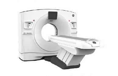
- GE’s unique Gemstone detector – lOOx foster aher glow time
- Powered by 100 KW generator, 8 MHU tube and 800 mA max current
- Ultra-fast kVp switching technology with perfect temporal registration for spectral imaging Unmatched spatial resolution in the industry of 0.23 mm
- Full SO cm Field of view whole body Dual energy application for anatomical coverage Prospectively gated cardiac scans with Spectral Imaging
- Upto 82% dose reduction using ASIR-V
Revolution CT-It's Time For A Revolution
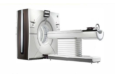
- High-definition imaging with the Gemstone TM Clarity Detector
- 160 mm coverage to achieve one-beat, motion-free coronary imaging at any heart rate
- Split second volume acquisitions to minimize motion artifact
- Volume Spectral CT – Faster than the blink of an eye
- GSI Xtream Workflow Makes Spectral CT Routine
- 437mm/s HyperDrive table speed for fast scanning acquisition
- 80 cm Wide bore to accommodate challenging patients including bariatric and trauma
- 25 msec Ultrafast kV switching technology for GSI Xtream
Revolution CT ES-Versatile and Exceptional CT Imaging
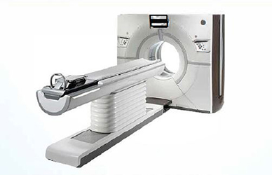
- High-definition imaging with the Gemstone TM Clarity Detector
- High coverage to achieve motion-free coronary imaging at any heart rate
- Split second volume acquisitions to minimize motion artifact
- Volume Spectral CT – Faster than the blink of an eye
- GSI Xtream Workflow Makes Spectral CT Routine
- 80 cm Wide bore to occommodate challenging patients including bariatric and trauma
- 25 msec Ultrafast kV switching technology for GSI Xtream
Optima 540-Your Diagnostic partner. Every Day.
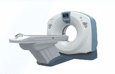
- Smarter option with state-of-the-art image quality
- Manage complex cases with smart technologies
- SmartMAR – single acquisition metal artifact reduction technology
- Clinical excellence through ASiR. IQ enhance and other applications
- Fast acquisition, reconstruction and post processing for improved workflow
- Increase operational efficiency upto 40% with Smart Flow
- Advanced visualization with AW multi modality workstation
Optima 520-Achieve Deeper Insight
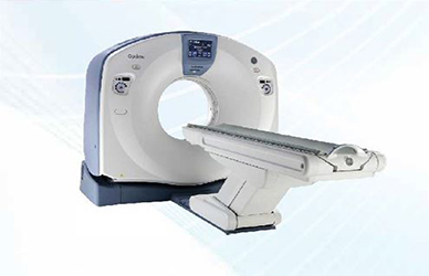
- Powerful intelligent CT with smart technologies
- SmartMAR – single acquisition metal artifact reduction technology
- Clinical excellence through ASiR, IQ enhance and other applications
- Fast acquisition, reconstruction and post prosessing for improved workflow
- Smart Flow improves efficiency and patient experience upto 40%
- Advanced visualization with AW multimodality workstotion
Discovery RT-see Everything. Miss Nothing.

- Comprehensive radiation therapy solution with precision. integration and efficiency
- Max DFOV of 80 cm feature provides edge-to-edge acquisition
- Enabled with Smart Metal Artifact Reduction Technology (Smart MAR)
- Powered by a lOOkW generator and 0.625 mm slice thickness
- Smart Deviceless 40 provides respiratory gating without an external device
- AdvantageSim MD enhonces productivity through effortless 4D review and simulation
Cath Lab
A “cath lab,” short for cardiac catheterization laboratory, is a specialized hospital room equipped with imaging technology to perform minimally invasive tests and procedures to diagnose and treat cardiovascular conditions using catheters.
Optima IGS 3 series-Cord,ovoscular Imaging Within Your Reach
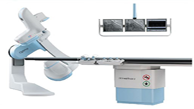
- Unique 3 Axis Gantry movement
- GE’s unique Stent Viz technology with subtracted guide wire
- Highest DQE – GE offers 80% at just 175 nGY
- Three Focal Spot (0.3 ,0.6 & 1.0mm, .3mm far DSA.)
- Intelligent Aut□Ex automatically selects the X-ray parameters
- Available in detector size of 20. cm x 20.5 cm and 30 cm x 30 cm
- In nova 3D provides o quick 3D rotational angiography acquisition
- GE Makes ifs detector at its own & is the only company unlike other vendors who get from otherOEMs
Innova IGS 5 series-Advanced V1suallzot1on And Multipurpose Appllcat,ons
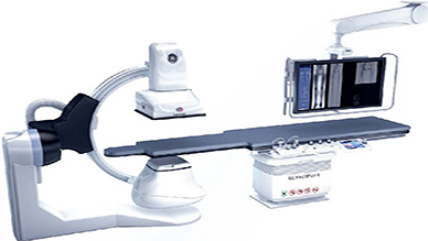
- AutoRight-GE’s unique and Industry’s first Al based imaging chain
- Ge’s self-made monolith detector
- GE’s unique Dose Blueprint Tech
- Unique 3 Axis Gantry movement
- GE Makes ifs detector at its own
- Highest DQE – GE offers 80% at just 175nGY
- Three Focal Spat (0.3 ,0.6 & 1.0mm, .3mm far DSA)
- 3DCT HD provides fine image details to help in clear visualizations
- Available in detector size of 20. cm x 20.5 cm, 30 cm x 30 cm & 40 cm x 40 cm
Innova IGS 6 series-v,ewTheAnotomyln Det01ls
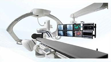
- AutoRight-GE’s unique and Industry’s first Al based imaging chain Unique offset C-Arm
- GE’s unique Dose Blueprint technology
- Highest DQE -GE offers 80% at just 175nGY
- One-hand full biplane control with the SmartBox
- C-arm design allows to perform 3D rotational acquisitions
- Low-dose imaging technologies far minimally invasive procedures Optimal detector size 120.5 x 20.5 cm 18.1 in), or 31 x31 cm square 112.2 in)
- Equipped with lnnovaSense, the advanced patient contouring technology
Discovery IGS 7 serieS-Redscover Space and Movement
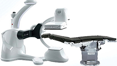
- AutoRight-GE’s unique and Industry’s first Al based imaging chain
- Robotic hybrid solution
- Wide bore offset C-arm
- Enables collision-free 2D & 3D imaging
- Lowest published dose levels far EVAR
- Unprecedented positioning versatility & full patient access from any side
- Longest table for long catheters & guide wires, surgical rail sleeve for OR accessories head-to-toe coverage from right/left side. No need for leg filters. Breeze subtracted bolus chase
- Planning anywhere, fusion guidance with Digital zoom & easy 3DCT HD assessment
- Customizable layouts and parking positions, which installs in even small rooms
Mammography
Mammography is an x-ray imaging method used to examine the breast for the early detection of cancer and other breast diseases. It is used as both a diagnostic and screening tool.
Senograp he CrystaI Nova-Digital Mammography Transformation, simphfied .
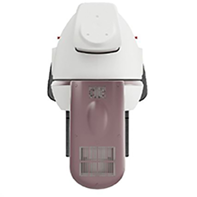
- Simple and intuitive features
- eContrast post-processing to optimize visual comfort
- Auto-Move function adapts to the height automatically
- High DQE detector from premium GE mammography systems
- N-button quickly and automatically positions workflow preferences
- Seno Iris Lite workstation integrated with the Senographe Crysta I Nova
- Small and sliding paddle makes it easy to accommodate all kind of patients
Senographe Pristina-Reshape The Mammography Experience
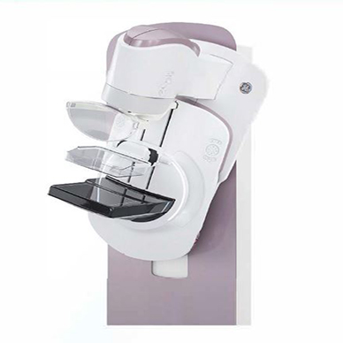
- Superior Diagnostic Accuracy
- Redesigned biopsy experience
- Exceptional 3D breast cancer screening technology
- Allows breast cancer diagnosis and treatment plan on same day
- SensorySuite distract patients from the perceived discomfort, pain and anxiety
Bone Mineral Density (BMD)
A Bone Mineral Density (BMD) machine, also known as a DXA (Dual-energy X-ray Absorptiometry) scanner, uses low-dose X-rays to measure bone density and assess bone strength, helping detect osteoporosis and predict fracture risk.
Prodigy advance-Bane and Metabolic Health
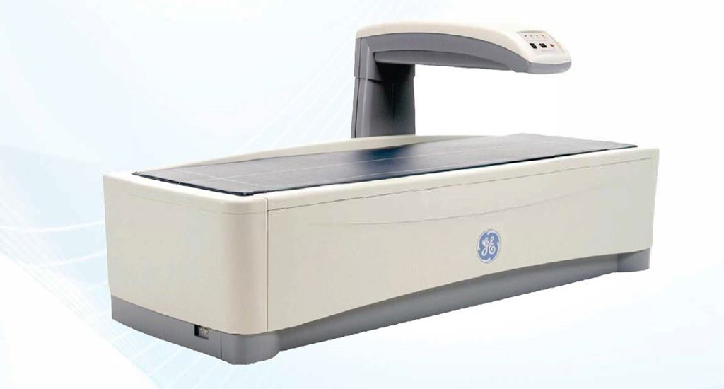
- Exceptional precision and low-dose radiation
- Streamlines patient care and practice workflow
- Provides precise data on soft tissue and bone composition
- Narrow-angle fan-beam with MVIR-that eliminates magnification error
- LYSO crystal detector provides efficiency and excellent energy resolution
- Efficiently provides accurate bone mineral density (BMD) and other body composition analyses
X-Ray
An X-ray machine uses X-rays, a form of electromagnetic radiation, to create images of the inside of the body, allowing doctors to visualize bones, fractures, and other internal structures for diagnosis and treatment.
Brivo XR115- Mobile-Ergonomically Designed For Added Safety
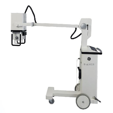
- Runs on lSAmps Electric Supply
- 200kHz UHF for low dose and Radiation
- <2 Kg Push force with small turning radius
- 4kW, lO0mA-Ultra High Frequency Mobile X-Roy
- 32GB Inbuilt Storage with Manual Stitching Software
- XR 120 Csl Detector with 127 micorns pixels & 70% DQE
Optima Traveller-Mobile-Takes Digital X-ray To The Point Of Care
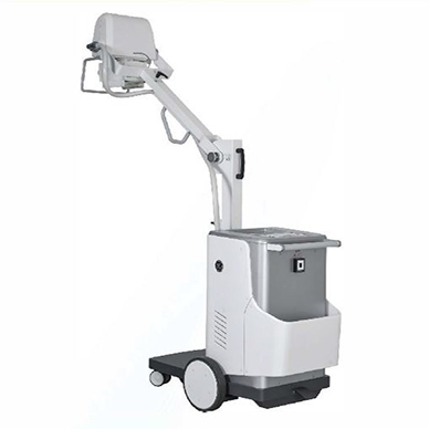
- APR Pre-looded-24 Techniques
- Non Motorized-Battery Free Operation
- Easy Maneuverability and Easy Positioning
- 32kW, 400mA-High Frequency Mobile X-Roy
- 32GB Inbuilt Storage with Manual Stitching Software
- XR 120 CSI Detector with 127 micorons pixels & 70% DQE
Optima XR 22Q-Opt1m1zed For The Woy You Work
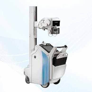
- 30kW High Frequency Generator
- Built for Reliability & Easy to Cleon
- On-board Integrated 15″ Touchscreen
- Fully integrated digital & Fully Motorized
- Stow While Charging-In Bin detector Charging Floshpod Detector-Non Tiled FPD with 68% DQE
Optima XR 240amx (Gen 2)-D1agnost1c Insight Close at Hond
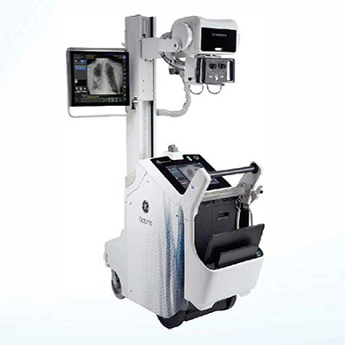
- RFID badge reader for foster access
- US FDA approved Critical Core Suite
- Remote connectivity for predictive maintenance
- HIS/RIS integration & AutoGrid for better workflow management Adaptive WiFi for better signal management while travelling across various hospital departments.
C-Arm
A C-arm machine is a mobile, flexible medical imaging device using X-ray technology to provide real-time, high-resolution images during surgical procedures, allowing surgeons to visualize internal structures and guide interventions.
OEC Brivo-Essental partner in Surgery
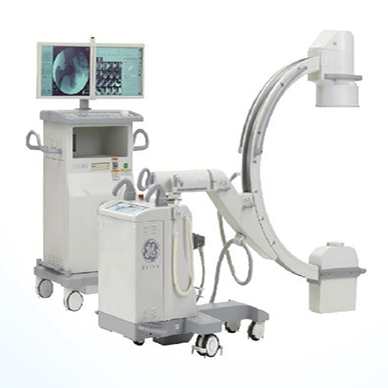
- Monochrome 19″ monitors
- Wireless DICOM and MPPS
- OEC interface allows confident operation
- lk x lk high resolution imaging technology
- 9″ Image Intensifier provides high spatial resolution
- CD/DVD, USB and integrated DICOM storage options Automated features, including true point-and-shoot capability
OEC One-Bringing you a Clear View of The lmages you Demand
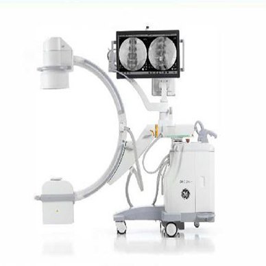
- 27″ image display on articulating arm
- Live image mirrored on TechView tablet
- Color-coded pivot joints and locks. along with laser aimers
- Wireless DICOM, digital video output and USB data transfer
- High res images with high DOE 165%1 image intensifier and lk x lk camera
OEC One CFD-See More. Do More
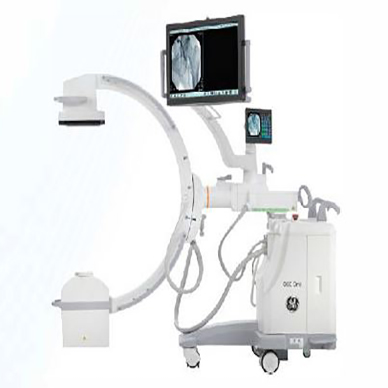
- 150,000 image storage
- View 4x image size with Live Zoom
- Small footprint and fits in tight spaces
- Standby power – 5-minute standby data protection
- 1:1 image detail with CMOS flat panel detector ICFD)
- Low dose mode and Finger-removable anti-scatter grid Synchronize workflow with simple commands for adjustments
OEC Elite II-Expect More
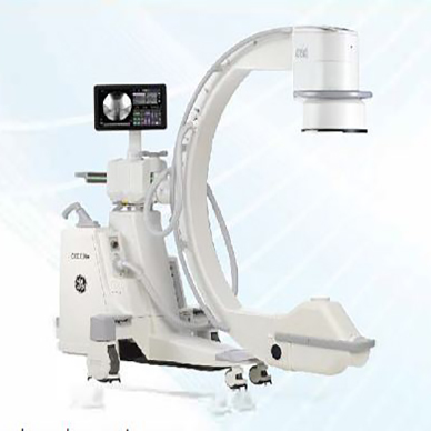
- Linux based operating system
- 4″ images 4K UHD 32″ display
- Intuitive OEC Touch control panel
- Lightweight slim design workstation
- Live Zoom, Digital Pen and measurement
- lK x lK x 16-bit image processing at 30 fps Disconnect and reconnect C-arm without rebooting
OEC Elite CFD-see What you've Been Missing
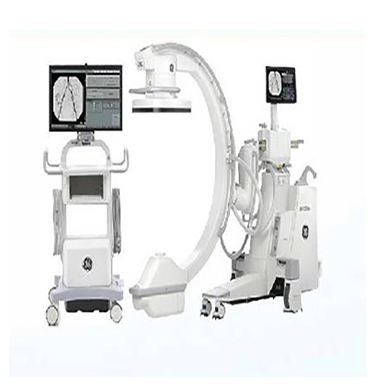
- 4K UHD 32″ display
- Intuitive OEC User interface
- Agile – 30% less force to steer
- Lightweight slim design workstation
- Efficient image capture with SmartView
- Live Zoom, Digital Pen and Measurement Largest Field of View IFOV)*see 20% more Continuous fluoroscopy allows to view more detail

